Table of Contents
Aim straight for your dog’s heart but it is not cupid but rather’ the filaria parasite. Filaria in dogs is a potentially deadly mosquito-borne disease that in its subcutaneous form can also affect humans.
Filaria is mainly prevalent in northern regions, particularly in lake areas, but cases of the disease are steadily increasing in other geographical areas of the South as well.
The gradual increase in temperature due to climate change means that mosquitoes are finding increasingly favorable conditions to spread, and the likelihood of infection and transmission of filaria is also increasing proportionally.
Existing forms of filaria
Depending on the parasite being carried, filariasis can have a cardiopulmonary form (caused by Dirofilaria immitis) or a cutaneous form (caused by Dirofilaria repens).
The most severe form is cardiopulmonary filariasis.
Usually the time of greatest danger is summer, because filaria requires at least 19 degrees to grow.
Cardiopulmonary filariasis generally affects large dogs living in an outdoor environment.
The disease develops silently because the parasite, before showing its presence, needs to grow and evolve.
The life cycle of cardiopulmonary filariasis larvae
A larva takes about 6-7 months before becoming an adult and remains infectious for years that’s why Filaria can occur even months after the time of infection and it is important to perform periodic testing at your veterinarian’s office to check your dog’s health status.
The mosquito bites an infected animal and ingests with blood the first-stage larvae, called microfilariae or L1.
Inside the mosquito the larvae mature, evolving from stage 2 (L2 larvae) to stage 3 (L3 larvae): this is the infesting form.
Having reached a sufficient level of growth to be re-injected, the microfilariae are transferred into the dermis of another dog, again through a puncture.
In the infested animal, the larvae migrate into the capillaries and within about 10 days transform into L4 larvae and later, progress to the L5 stage.
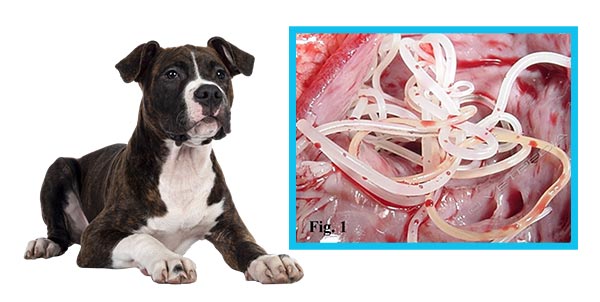
The larvae, once they become adult worms, settle in the heart, particularly in the atrium and right ventricle and lungs.
The adult parasites, called macrofilariae, grow to a considerable size: the male averages 12 to 20 cm in size, while the female reaches up to 30 cm.
They grow and spread so widely that if the disease is not diagnosed and treated in time, it can lead to your dog’s death.
Cutaneous filariasis
Adult worms of Dirofilaria Repens larvae, on the other hand, go to localize in the subcutis, causing significantly less damage.
Clinical signs induced by adult filariae are subcutaneous nodules containing filariae.
These nodules are mobile, soft, and have serum-hemorrhagic content.
They range in size from 3-6 cm and in most cases can be confused with tumor formations for which surgical removal is indicated.
Other symptoms are alopecia, generalized dermatitis, itching and granulomas.
What symptoms do dogs with cardiopulmonary filariasis exhibit
Dogs affected by filariasis may exhibit symptoms that will be as severe as the number of parasites in the body.
However, the first symptoms appear several months after infestation, when the body reacts against the parasite and the heart, damaged and exhausted by the presence of filariae, begins to function poorly.
The dog appears tired, coughs, is fatigued even after minor exertion, and, as time passes, becomes increasingly inappetent and depressed.
Thereafter, the cardiovascular damage worsens and the disease negatively affects the whole body, causing breathing difficulties, fluid accumulation in the abdomen and even neurological problems.
What diagnostic tests reveal the presence of heartworm in dogs
To test for the presence of filaria, it is sufficient to submit the dog to a blood test , which, given the particularity of the disease and the timing of incubation, is useful to repeat annually.
This test should be performed in the spring in all individuals living in endemic areas.
Once the presence of the parasite in the circulation is established, it is of paramount importance to understand where it has localized through more extensive examinations: chest X-rays show the health of veins and pulmonary arteries, and echocardiography allows assessment of the effects of filaria on the heart.
Treatment for filariasis
Treatment is complex and involves drugs that must be carefully dosed by the veterinarian according to the stage of the disease and the dog’s overall health.
The veterinarian usually proceeds with what is known as adulticide therapy, a treatment that allows the killing of adult filariae that have settled in the heart and lungs.
In the more advanced stage of the disease, in addition to symptomatic therapy, surgery to remove macrofilariae is often necessary.
If heartworms have been in the dog’s heart for a long time, the dog is likely to remain cardiopathic even after treatment, as the damage caused to the heart is not always reversible.
How to take preventive action against filaria
The most effective prevention is simultaneously repellent and abatement and must be carried out to achieve two main objectives:
-Reducethe possibility of being bitten by the vector: special repellents are commercially available to prevent mosquitoes from coming into contact with your dog.
-eliminatelarvae that may have been injected anyway: to inhibit the development of adult parasites, give your dog a microphilarial therapy.
This treatment prevents larvae development from the L3 and L4 forms to the adult form in the infested dog.
Ivermectin is the most widely used active ingredient to simultaneously prevent both cardiopulmonary filariasis and subcutaneous filariasis: it eliminates mosquito-inoculated larvae in the 30-40 days prior to treatment, intervening before they begin their migration to the heart.
Contact the Veterinary Doctors on our staff for preventive, routine, and specialty examinations and diagnostic tests to be performed on-site.
Clinica La Veterinaria is always open h24 every day including holidays and with First Aid service from 8 pm to 8 am.
For the joy of seeing them HAPPY.


