
Diagnostic imaging
Diagnostic imaging:
X-rays, ultrasound and echocardiography for complete and accurate diagnosis in dogs and cats of all sizes and ages
Imaging is a painless, noninvasive, repeatable method that almost never requires anesthesia.
Radiology, the true cornerstone of diagnostics, is mainly applied to study the skeletal system but also to explore the chest and abdomen of dogs and cats.


The X-ray system we use is direct digital and has many advantages over the traditional film technique:
Ultrasound is also an essential technique in veterinary imaging. It allows very detailed images of the internal organs of dogs and cats to be obtained in real time and their morphological and structural changes to be assessed.
Ultrasound is an indispensable examination:
Echocardiography allows us to visualize the heart in motion (real-time), take measurements of cardiac contractility, assess wall thickness, flow dynamics, and valve integrity.


For X-rays, ultrasound and echocardiography, we employ specialized personnel.
In case of an emergency in the emergency room, if the symptomatology involves the chest and abdomen, it is always possible to subject the dog or cat to Fast Echo.
Read these articles

Kidney stones and urinary stones in dogs and cats: a serious and very dangerous emergency. Here are the symptoms and precautions to take.
The presence of stones in the kidneys and urinary tract of dogs and cats is a serious emergency that can even prove fatal.
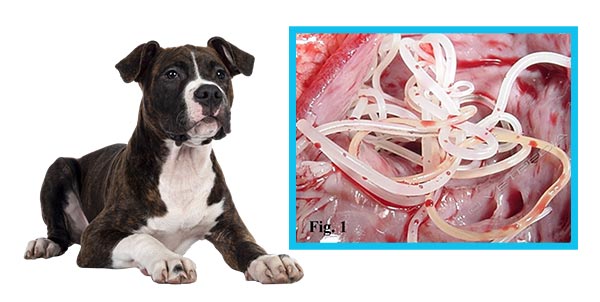
The heartworm parasite silently strikes your dog’s heart and puts it in serious danger of death. How to prevent this from happening?
Filaria or filariasis: increasingly common but little known, it can silently kill your dog. Prevention first.