Mastocytoma in dogs is a very common tumor: it presents as a cutaneous or subcutaneous nodule and accounts for about 30% of all neoplasms. How to recognize a cutaneous mastocytoma in the dog?
Mastocytoma is manifested by the appearance of a well-circumscribed, hard, raised, hairless, cutaneous or subcutaneous nodule that varies in size from about 0.5 to 2 to 3 cm and tends to grow more or less rapidly.
If the nodule is ulcerated, it causes the dog irritation to the point of scratching to the point of injury.
The prevalent location is in the trunk, perineal area, on limbs, head and neck of the dog.
The underlying causes of mastocytoma
Mastocytoma is caused by hyper proliferation of mast cells (MCs), naturally occurring immune system cells deputed to activate defensive responses against microorganisms and other foreign molecules.
The causes associated with the development of mastocytoma remain difficult to assess, and it is leaning toward a multifactorial approach where genetic predisposition assumes a major role.
The most accepted theory is a gene mutation in mast cells, particularly the C-Kit gene.
Mutated mast cells lose their normal functions and degenerate into neoplastic cells.
In fact, mastocytoma in dogs can affect any breed and more or less young subjects.
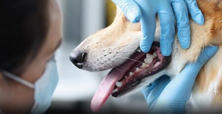
Diagnosis: the cytological examination
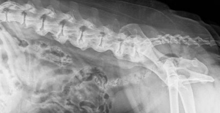
The veterinarian will perform chest X-rays, abdominal ultrasound, and cytological examination of spleen, liver, and relevant lymph nodes through needle aspiration.
By looking at the cells of the sampled material under the microscope, the Veterinarian will be able to determine whether it is mastocytoma or something else such as a papilloma, for example.
Once mastocytoma is diagnosed, staging should be performed
Some mastocytomas in dogs behave indolently even for years; others are more aggressive and metastasize quickly.
Staging is necessary to assess the invasiveness and spread of the neoplasm and allow the Veterinarian to evaluate the most appropriate therapeutic treatment for the dog.
Basically, it has to be ascertained whether there is widespread metastasis to internal organs (liver, spleen) and whether the tumor has already affected the lymph nodes.
After the cytological examination, if necessary, The Veterinarian will proceed with the histological examination, which consists of taking small portions of liver and spleen tissue.
Therapy and treatments: how to treat a dog with mastocytoma
Surgery is the therapy of choice for most mastocytomas that have not metastasized, or have metastasized to the regional lymph node.
The regional lymph node is the lymph node that drains the lymph from that particular body region of the dog.
In addition to removal of the primary tumor, it is also always useful to remove the sentinel lymph node on which histologic examination and staging will be performed.
If surgery is not possible, for example when the nodules are multiple and not completely removable or the neoplasm has already metastasized, chemotherapy is necessary.
Prognosis
The prognosis varies greatly depending on the histologic grade of mastocytoma, the status of the sentinel lymph node, and the staging result.
One thing is certain: the timeliness of diagnosis matters.
Any suspicious neoformation or new skin lesion that shows a tendency to grow and change shape found on your dog should always be immediately investigated by your Veterinarian.
To refer your dog for a checkup, contact the veterinary doctors on our staff who are always available to you.
We would also like to remind you that Clinica La Veterinaria is always open h24 every day including holidays and with First Aid service from 8 pm to 8 am.